A couple posts ago, we began to talk about what is involved in a “simple” cat spay  operation. If you missed it, you can catch up now to learn more about the steps leading up to a spay. We left off at the point where the surgeon has located one horn of the uterus.
The surgeon, using the uterus as a guide, traces “up” to where the ovary is attached to the end of the uterus. This entire structure is then attached to the interior body wall by a short fibrous band called the suspensory ligament. In order for you to get a mental picture of what all this looks like and how it is put together, realize that the most experienced veterinary surgeons perform this surgery through an incision in the body wall that is between one half and one inch long! The uterus is a tube about the diameter of a Bic pen refill, but it can swell and thicken to about the thickness of a pencil when the cat is in heat. The ovary is about a half inch in length, and the suspensory ligament is sometimes less than that. In order to get the ovary out of the body, the surgeon has to carefully pull on this ligament to stretch it enough to pull the ovary up out of the abdomen from its usual location high in the back.
Once the ovary has been exteriorized (pulled up out of the abdomen), the artery and vein that supplies it, and that run parallel to the suspensory ligament, must be ligated (tied off) to prevent bleeding after the ovary is cut loose. Usually one or two clamps are first placed between the ovary and the body and the ligature then applied between the two or over where one of these clamps was placed after the surgeon removes it. Most ligatures are made of a sterile absorbable material, but some veterinarians use metal clips or even stainless steel suture to tie things off.
When the surgeon is confident that the ligature(s) are secure and the blood vessels won’t begin to bleed, the cut end of the ovarian artery/vein pedicle is gently released to pull back into the abdomen.
Now the surgeon traces back down the “arm of the Y” to where the two arms join at the body of the uterus and finds the other uterine horn that runs up to the other ovary. The same procedure is performed to exteriorize it from the abdomen, ligate the vessels, and cut the ovary and uterine horn free.
In technical terminology, a spay operation is called an ovariohysterectomy. This means that both ovaries and the entire uterus are removed. You want to remove the uterus and both ovaries for a number of reasons. If you don’t remove the ovaries, the cat will continue to go into heat, howling and rolling on the floor for weeks a couple of times a year. So it’s pretty obvious why we want them out, but why remove the uterus? We do this because, once the ovaries are out, the uterus becomes both a useless organ and poses a risk to the cat. The uterus is a hollow tube that can now become infected. If a pyometra (infected uterus) develops, the cat will become very ill and could die.
Now with both ovarian arteries and veins ligated and cut, the surgeon moves down to where the horns meet to form the body of the uterus, the “stem of the Y.” Ligatures are placed around the base and the uterus is cut free and removed. Again for reference, this part of the reproductive tract is located low in the abdomen near the pelvis between the bladder and the colon. In most cats, this is three to four inches away from where the ovaries attach.
 The cut in the linea alba is closed with sutures. Some surgeons use an absorbable material so that the stitches will dissolve in time, while others prefer to use a non-absorbable material that will remain relatively inert. Depending on the size of the incision and other factors, the surgeon may place additional sutures in the tissues below the skin to pull the incision together. Finally, the skin incision is closed. If needed, stitches are placed in the skin or the incision is glued closed.
But we’re not done yet. The cat still has to recover enough from the anesthesia to be able to regain protective reflexes such as the ability to swallow. A veterinary technician or nurse must monitor the patient as she wakes up until this reflex returns. When it does, the endotracheal tube is removed and the cat is allowed to rest and recover.
Later that day, you get to come by and bring the little girl home. In most cases, the cat gets to eat some dinner and sleep off the rest of the effects of the anesthesia. By the next day, she should be up and around asking for some good petting and attention. If there are any skin sutures, these are removed in a week or two.
This blog post was adapted from a newsletter article originally written by Dr. René Gandolfi of Castro Valley Companion Animal Hospital.


 1. Always give generously. A small bird or rodent left on the bed tells them you care.
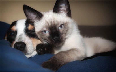
 me, it is 16 years and up. Some people are afraid to adopt an older cat because they fear losing their beloved new family member too soon. Of course, there are no guarantees in life, but cats generally live much longer than most dog breeds, especially Siamese cats. Cats over 20 are not hard to find. With appropriate healthcare, just as with humans, animals are getting older. If you think about it a certain way, adopting a 10-year-old cat is not much different than adopting a large-breed puppy.
 People often think it is easier to integrate a kitten and that an older cat may come with past baggage, making it difficult to adjust quickly. Usually, the opposite is true. Like many shelter animals, cats just want to belong. A cat growing up with amenities knows a good thing when she sees it and really wants to be a part of it. Older cats also know all about litter boxes and do not need a lot of training. They forgive you if you work late and will not be waiting to greet you from atop the curtain rod.


 Further enforce this by feeding the animals near each other, but with a door separating them. This will help them associate the other’s scent with the positive result of being fed.
 break up any conflict. Do what you have to do to get your animals separated and try again later. If this continues to happen, you might need to get an animal behaviorist involved.


 My cat is over 10 years old. Is there anything I should be doing for her to help maintain optimum health?